

The flatfoot
Idiopathic flatfoot is characterized by a lowering of the arch on the inner side of the foot. In children under three years, flat feet are normal due to abundant fatty tissue development. Initially, there is a physiological flatfoot condition related to relative muscle insufficiency and ligament laxity typical of early childhood. As the child grows, bone morphology changes through the absorption of adipose tissue, strengthening of the plantar muscles, and reduction of ligament laxity. Generally, the foot acquires normal shape and function around 6-7 years of age.
CAUSES
The causes of flat feet can vary:
- Congenital (fusion between foot bones, known as synostosis, complex malformative syndromes, short Achilles tendon)
- Neuromuscular (secondary to diseases of the nervous and muscular systems such as Cerebral Palsy, Myopathies, Spina Bifida)
- Post-traumatic (calcaneus fracture, astragalus fracture)
- Inflammatory (juvenile idiopathic arthritis)
- Idiopathic (unknown cause)
SYMPTOMS
The most common form is idiopathic valgus flatfoot in childhood, and its etiology is still unknown, although it is often associated with family history, ligament laxity, and reduced tone of the arch muscles. Idiopathic flatfoot is more common in overweight children and adolescents. In pediatric cases, flat feet are generally asymptomatic; however, around 6-7 years of age, children may begin to experience fatigue and leg pain during walking and physical activities.
EVALUATION CRITERIA
Another crucial clinical assessment to decide on the type of treatment is distinguishing between the flexibility and rigidity of flat feet. Flexibility, the correctability of flat feet, is evaluated with various clinical tests:
- Observation of the foot under load: If the arch is absent under load but appears when unloaded, the foot is flexible.
- Tip-toe test: Observation under load and then raising onto tiptoes; if the arch reappears, the flat foot is flexible.
- Jack test: Passive bending of the big toe under load; if the arch reappears, the flat foot is flexible.
Flexible asymptomatic flat feet may have a favorable spontaneous evolution and respond well to conservative treatment. Rigid flat feet are often symptomatic and less responsive to conservative treatment. Flat feet typically exhibit medial support (pronation) and lateral tilting of the heel (calcaneal valgus).


DIAGNOSIS
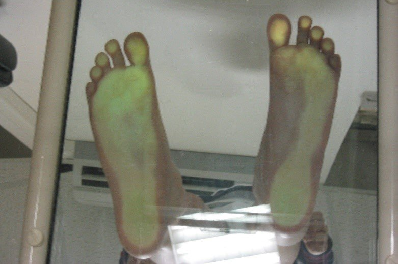
The diagnosis of flat foot is fundamentally clinical, the podoscope is an examination that shows the shape of the foot under load very well by showing the plantar footprint through an illuminated mirror. (SEE PHOTO)
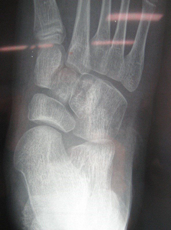
The radiographic examination under load allows to evaluate the physiological angles of the foot (Kite angle, Costa-Bertani angle). In cases of congenital malformations (synostosis), an evaluation through CT scan or MRI is appropriate.
FLAT FOOT ON THE RIGHT, NORMAL FOOT ON THE LEFT
TREATMENT
The treatment of flat feet is still a matter of discussion among different schools of thought. Initially considered an aesthetic deformity, if not corrected spontaneously or with orthoses' help, it can cause functional symptoms (fatigue, pain) due to improper load distribution, muscle imbalances, and altered joint biomechanics. Conservative treatment includes physical therapy (rehabilitative exercises) and orthoses (insoles). Although subject to conflicting opinions, it can be useful to provide biomechanical support to the foot through insoles and strengthen the arch muscles through specific exercises like walking on tiptoes and heels, and walking on sand during the summer.
Surgical treatment is indicated in non-physiological flexible flat feet (pain or progressive worsening) and symptomatic rigid ones. Surgery is performed only in cases without favorable evolution by 8-14 years and where pathological pronation persists in all phases of walking, thus at risk of secondary degenerative pathology in adolescence or adulthood. These selected cases, with an unfavorable prognosis, constitute no more than 2-5% of all clinically observed flatfoot cases in the developmental age.
SURGICAL TECHNIQUE
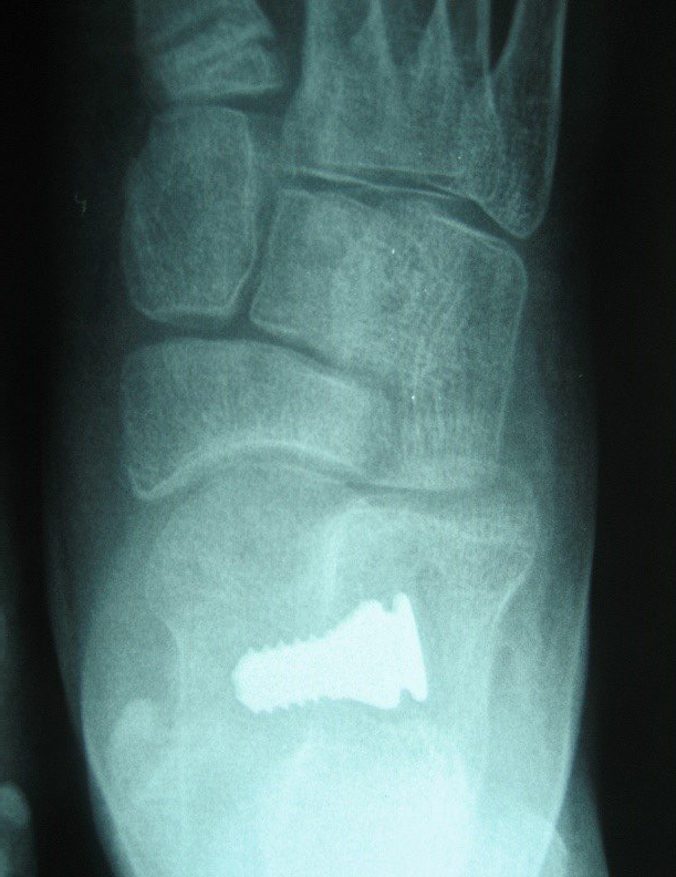
The preferred surgical intervention for idiopathic flat feet in the developmental age is subastragalar arthrodesis (the space between the talus and calcaneus), performed between 8 and 14 years. Arthrodesis refers to an operation that limits the movement of a joint without causing permanent ankylosis (loss of movement). Arthrodesis involves applying corrective screws, which can be made of metal (titanium or steel) or absorbable material (poly-L-lactic acid). Non-absorbable endorteses are usually removed at the end of growth, while absorbable ones have the advantage of not requiring removal but entail a higher risk of deformity recurrence.
TITANIUM ENDORTESIS

ACCOMMODATION OF THE ENDORTHESIS IN THE SINUS TARSOUS (SPACE BETWEEN THE TASTALUS AND THE CALCANUS)

SURGICAL PROCEDURE
The surgical incision is very small, about 2 centimeters, and the surgical and anesthesiological times are very short, approximately 15 minutes per foot. Using a guide wire under fluoroscopic control, the endortesis is inserted into the tarsal sinus (the space between the talus and calcaneus). Recovery times are rapid, with no plaster casts applied. In the first postoperative week, the patient remains non-weight-bearing and begins active foot mobilization exercises. After one week, weight-bearing is introduced with the help of Canadian crutches, which are abandoned after about two weeks.
Arthrodesis not only exerts a mechanical action by correcting the relationship between the talus and calcaneus but also plays a neuromotor role as it is implanted in a foot area rich in receptive nerve endings capable of activating supinator muscles. In a growing subject, the restoration of anatomical relationships between foot bones obtained with surgical intervention leads to bone restructuring and adaptation of soft tissues over time, stabilizing the correction even after the endortesis is removed or absorbed.
LATERAL PRE-OPERATIVE RADIOGRAPHIC PICTURE

LATERAL POST-OPERATIVE RADIOGRAPHIC PICTURE

PRE-OP IN ANTERO POSTERIOR
POST-OP IN ANTERO POSTERIOR
written by Pier Francesco Costici
Bambino Gesù Children Hospital (Rome)
original article from ospedalebambinogesù.it